Treatment of Colonic Volvulus and Acute Colonic Pseudo-Obstruction (Ogilvie’s Syndrome)
Colon obstructions can be mechanical or non-mechanical and constitute about 25% of all intestinal obstructions. Among the mechanical causes, the most common are:
- obstructive tumor in the colon or rectum (60%);
- stenosis due to previous diverticulitis (10%);
- volvulus of the colon (15 to 20%).
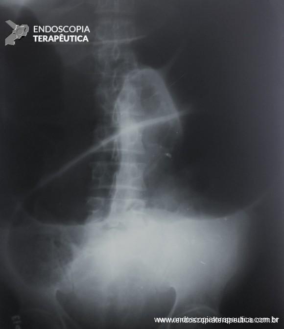
Colon volvulus is the twisting of a redundant segment of the colon on its mesentery that can lead to luminal occlusion of the twisted segment and ischemia by rotation of the mesocolon and, consequently, to perforation.
Although colon volvulus can occur in any redundant segment, it most commonly involves the sigmoid (60%–75% of all cases) and cecum (25%–40% of all cases).
Sigmoid volvulus occurs mainly during the 6th to 8th decades of life, being more common in men, institutionalized patients, patients with chronic constipation, neuropsychological impairment, or decompensated comorbidities. On the other hand, cecal volvulus usually presents in younger patients and has a female predominance.
Acute pseudo-obstruction of the colon, or Ogilvie’s syndrome, is a non-mechanical functional cause of obstruction that is believed to be a consequence of dysregulation of the autonomic impulses of the colon’s innervation. There is significant distension of the colon without an obstructive factor, but it can also evolve into ischemia and perforation. Clinical presentations vary according to the degree of distension, whether the ileocecal valve is competent or not, and the clinical condition of the patient. More commonly, Ogilvie’s syndrome affects elderly patients or patients hospitalized for unrelated reasons, including elective surgery, trauma, or treatment of an acute medical condition.
Here we present some recommendations from the guidelines of the American Society of Colon and Rectal Surgeons for the management of these cases.
Colon Volvulus
- Initial evaluation with history, physical examination, and basic laboratory tests. Symptoms may include cramping, nausea, vomiting, abdominal discomfort. Sigmoid volvulus usually has a more indolent presentation, while cecal volvulus tends to have a more acute presentation. On physical examination, there is generally abdominal distension with varying degrees of tenderness to palpation, up to peritonitis. Rectal examination reveals an empty rectal ampulla. Presentation in the emergency room with peritonitis and signs of shock occurs in 25 to 35% of cases.
- In hemodynamically stable patients, an abdominal radiograph aids in the initial evaluation (finding of “coffee bean” and, in patients with incompetent ileocecal valve, small bowel distension). Computed tomography is used to confirm the diagnosis.

Sigmoid Volvulus
- Hemodynamically stable patients without signs of peritonitis or evidence of perforation should undergo rectosigmoidoscopy to assess the viability of the sigmoid, untwist the torsion, and decompress the colon, effective therapy in 60 to 95% of cases. It is possible to maintain a decompression tube after rectosigmoidoscopy. The recurrence rate is 43 to 75% in cases not subjected to subsequent surgical intervention.
- Urgent sigmoidectomy is indicated when endoscopic detorsion is unsuccessful and in cases of colon suffering or perforation, as well as in patients with signs of peritonitis or septic shock. After resection of the twisted segment, the decision to perform a primary anastomosis, terminal colostomy, or anastomosis with diversion should be individualized considering the clinical context of the patient at the time of surgery, the conditions of the remaining colon, and comorbidities.
- Patients who undergo successful endoscopic detorsion are candidates for segmental colectomy during the same hospital admission to prevent recurrent volvulus and its complications. Non-resection operations, including only detorsion, sigmoidopexy, and mesosigmoidoplasty, are inferior to colectomy for the prevention of recurrent volvulus.
- Endoscopic fixation of the sigmoid may be considered in selected patients in whom surgical intervention is prohibitively risky.

Cecal Volvulus
- Attempts at endoscopic reduction of cecal volvulus are not recommended.
- Segmental resection is the treatment of choice for patients with cecal volvulus. Nonviable or ischemic cecum is present in 18% to 44% of patients with cecal volvulus and is associated with a significant mortality rate.
- In the case of cecal volvulus with viable intestine, the use of non-resection surgical procedures should be limited to patients without clinical conditions for resection.
Acute Colonic Pseudo-Obstruction (Ogilvie’s Syndrome)
- The initial evaluation should include history and physical examination, laboratory tests, and imaging diagnosis.
In the absence of fever, leukocytosis, peritonitis, pneumoperitoneum, or cecal diameter > 12 cm, initial therapy consists of correcting hydroelectrolytic disorders, volume replacement, avoiding or minimizing the use of opioids, avoiding anticholinergic medications, and identifying and treating concomitant infections. Deambulation, fasting, positioning maneuvers (knee-chest or prone) to promote intestinal motility, and decompression with nasogastric and rectal tubes are also recommended. Oral osmotic laxatives should be avoided as they can worsen colon dilation. Abdominal radiographs are part of the daily evaluation, accompanied by physical examination.
- The initial treatment is clinical support and includes the exclusion or correction of conditions that predispose patients to the condition or prolong its course.
- Pharmacological treatment with neostigmine is indicated when the condition does not resolve with supportive therapy.
- Endoscopic decompression of the colon should be considered in patients with Ogilvie’s syndrome in whom neostigmine therapy is contraindicated or ineffective.
- Surgical treatment is recommended in cases complicated by ischemia or perforation of the colon or refractory to pharmacological and endoscopic therapies.
References:
- Alavi K, Poylin V, Davids JS, Patel SV, Felder S, Valente MA, Paquette IM, Feingold DL; Prepared on behalf of the Clinical Practice Guidelines Committee of the American Society of Colon and Rectal Surgeons. The American Society of Colon and Rectal Surgeons Clinical Practice Guidelines for the Management of Colonic Volvulus and Acute Colonic Pseudo-Obstruction. Dis Colon Rectum. 2021 Sep 1;64(9):1046-1057. doi: 10.1097/DCR.0000000000002159. PMID: 34016826.
- Yeo HL, Lee SW. Colorectal emergencies: review and controversies in the management of large bowel obstruction. J Gastrointest Surg. 2013;17:2007–2012.
- Bauman ZM, Evans CH. Volvulus. Surg Clin North Am. 2018;98:973–993.
- Quénéhervé L, Dagouat C, Le Rhun M, et al. Outcomes of first-line endoscopic management for patients with sigmoid volvulus. Dig Liver Dis. 2019;51:386–390.
How to cite this article
Camargo MGM. Treatment of Colonic Volvulus and Acute Colonic Pseudo-Obstruction (Ogilvie’s Syndrome). Endoscopy News, 2024, vol1. Available at: https://endoscopy.news/2024/02/05/treatment-of-colonic-volvulus-and-acute-colonic-pseudo-obstruction-ogilvies-syndrome/