Role of Endoscopic Ultrasound in Idiopathic Acute Pancreatitis

Introduction

Acute pancreatitis is one of the diseases responsible for the highest number of emergency hospitalizations in gastroenterology. Its incidence is 13 to 45 cases per 100,000 people, accounting for 270,000 hospitalizations per year in the United States 1. Most cases are mild, but it can evolve into severe forms, requiring intensive care unit admission and even death. Its mortality is close to 5%, being significantly higher when only the most severe cases are analyzed 2.

Gallstone disease and alcoholism are the main causative agents, accounting for about 60% to 80% of cases 3. Other less common causes include anatomical changes, metabolic disorders, tumors, autoimmune diseases, among others. However, in a significant percentage of cases, about 10% to 30%, it is not possible to identify a causative factor after initial evaluation 3-4. It is then defined as idiopathic acute pancreatitis, being the 3rd most common cause in some series 3.

A detailed evaluation in these patients with idiopathic acute pancreatitis is of fundamental importance, since 14% to 26% may present recurrent episodes, evolving to chronic pancreatitis 5-6. In some cases, after specialized examinations, a treatable causative agent can be identified, thus avoiding new crises.

The endoscopic ultrasound is a minimally invasive procedure and, due to the proximity of the stomach and duodenum to the pancreas and bile ducts, allows a detailed examination of this region. Several studies have shown its benefit in the investigation of patients with idiopathic acute pancreatitis, however, the timing of its performance is not yet well defined 1,2-8.

Accuracy of Endoscopic Ultrasound

The accuracy of endoscopic ultrasound in identifying a causative agent in patients with idiopathic acute pancreatitis varies greatly among studies, from 29% to 88% 4. This large difference is due to the inclusion criteria used in each study. In those where patients underwent a greater number of diagnostic tests before the endoscopic ultrasound, the accuracy was lower. In those where patients were referred earlier for endoscopic ultrasound, the accuracy was higher.

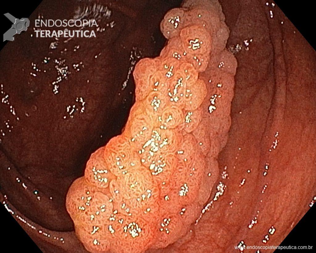
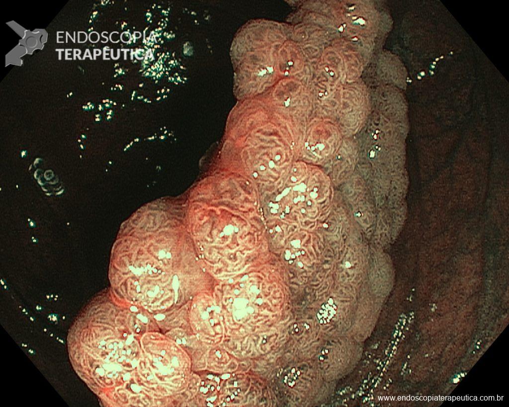
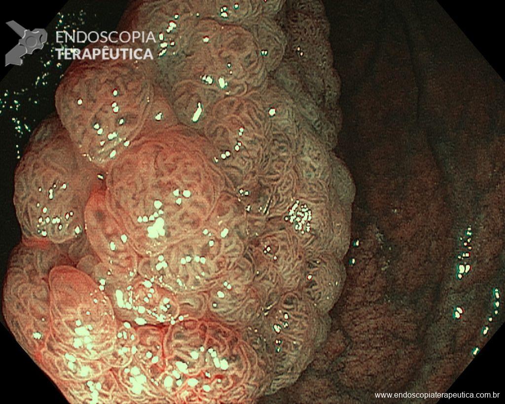
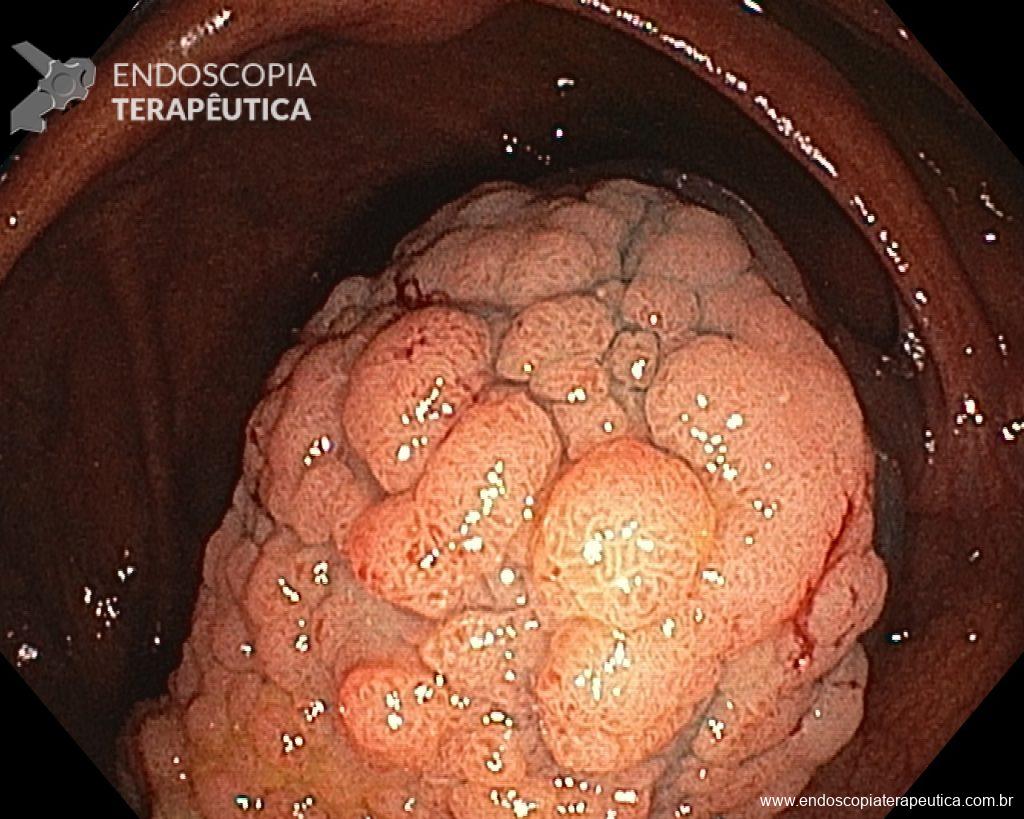
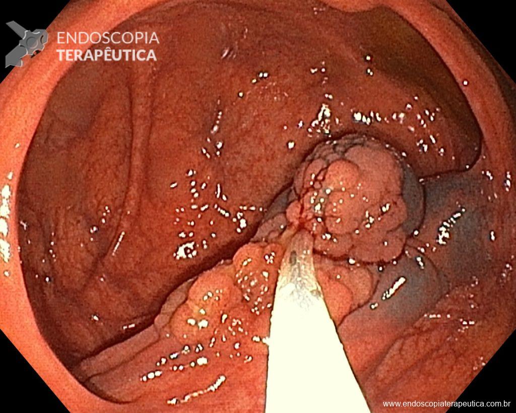
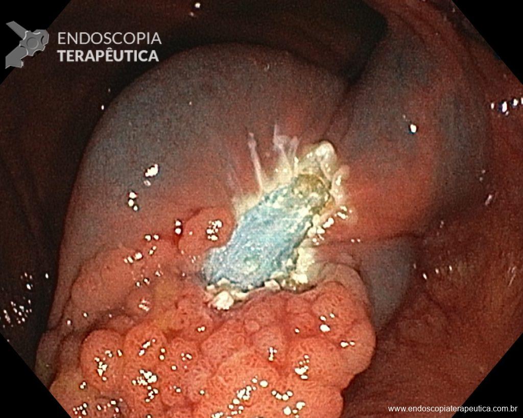
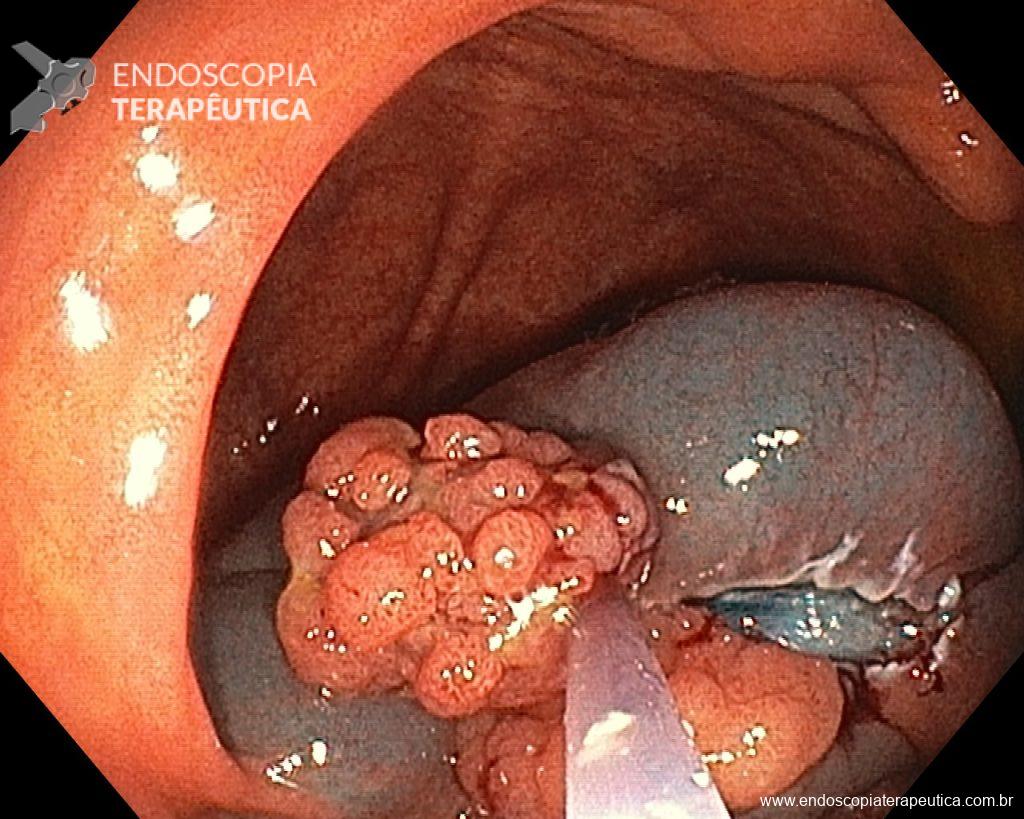
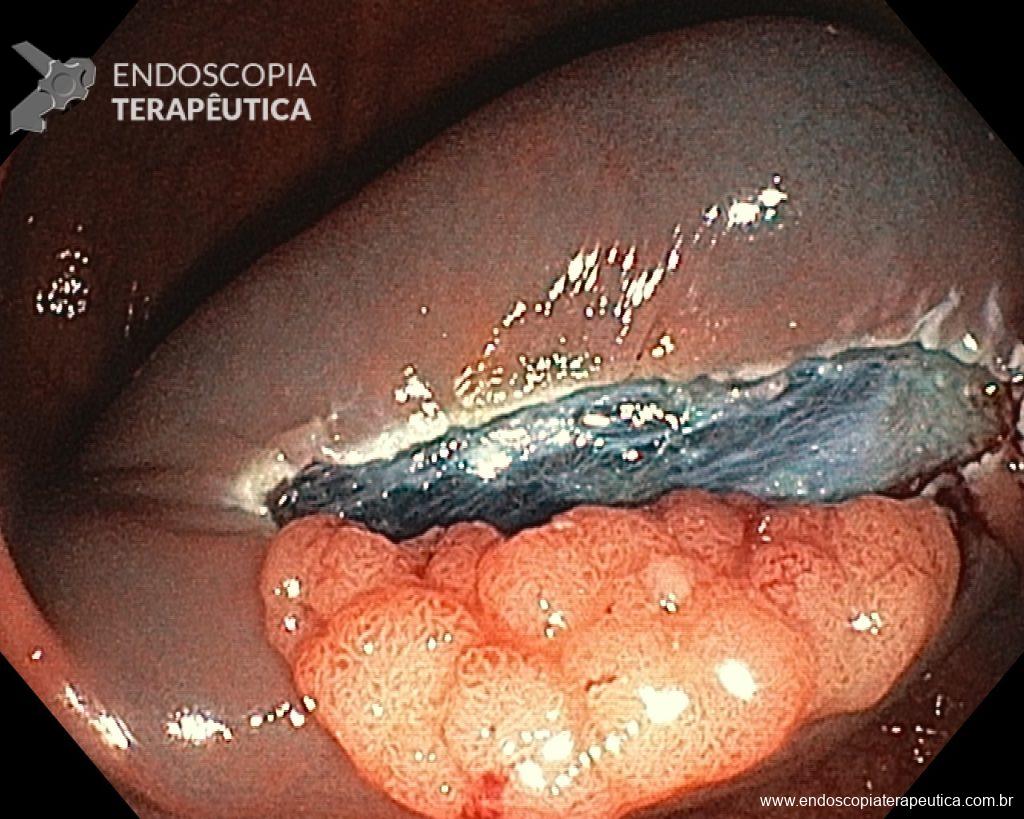
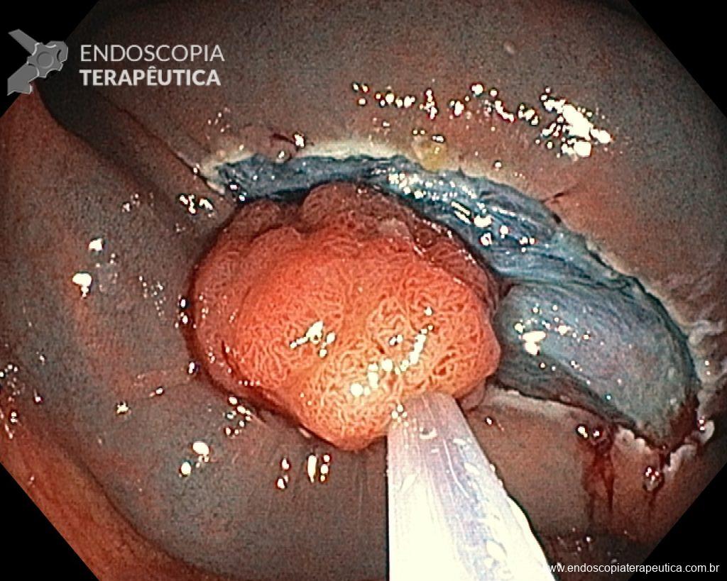
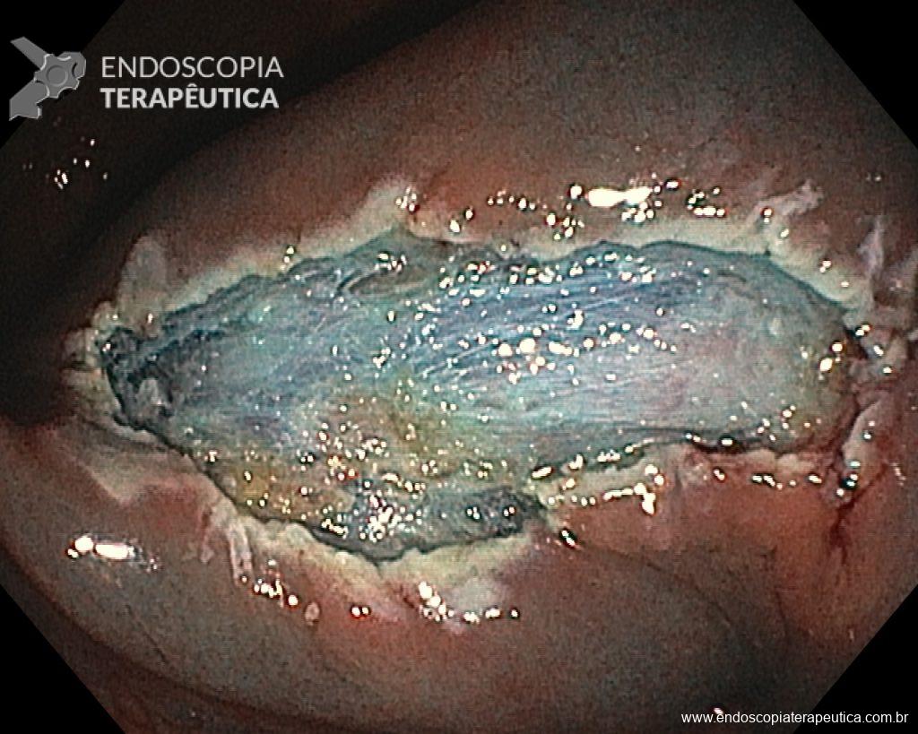
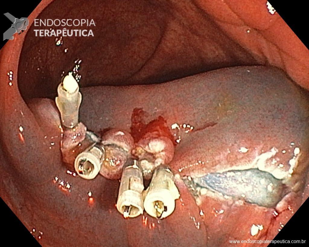
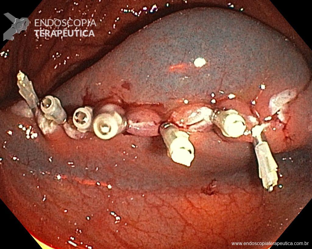
Umans and colleagues in a recent meta-analysis reached an accuracy of 59% 7. Biliary lithiasis, presence of stones, microlithiasis or biliary sludge in the gallbladder or common bile duct, was the most common cause, accounting for 30% of cases (Figures 1 and 2). In second place came chronic pancreatitis with 12% and in third pancreas divisum with 5%. It is important to note that in 2% of patients a neoplasm that had not been diagnosed in previous exams was detected. The identified lesions were intraductal papillary mucinous neoplasms (IPMN), pancreatic carcinomas, neuroendocrine tumors (Figure 3), adenomas, and papillary carcinomas. Other less common causes were autoimmune pancreatitis, ascariasis, choledochocele (Figures 4 and 5), biliopancreatic junction anomaly, and diverticulum.

In the same meta-analysis, when comparing the accuracy of endoscopic ultrasound in patients who had previously undergone cholecystectomy with those who had not, the result was different between the two groups, being 50% and 64%, respectively 7. Thus demonstrating how gallbladder lithiasis is indeed one of the most common causes.

When to Perform Endoscopic Ultrasound After an Episode of Pancreatitis?

There is controversy in the literature about the ideal timing for endoscopic ultrasound after an episode of acute pancreatitis 5. Authors who suggest performing the procedure earlier, sometimes while the patient is still hospitalized, argue that a possible diagnosis could be made more quickly, avoiding the possibility of recurrence and also preventing the patient from losing follow-up 5. Those who prefer to perform the procedure later, about 4 weeks after the resolution of the case, argue that the inflammatory changes secondary to pancreatitis could complicate the diagnosis, reducing the accuracy of endoscopic ultrasound 5.

In the meta-analysis by Umans and colleagues, the accuracy of endoscopic ultrasound after the improvement of acute pancreatitis and before improvement was 61% and 48%, respectively 7.

Perform Endoscopic Ultrasound After the First Episode of Idiopathic Acute Pancreatitis or Only in Recurrent Cases?

There is no consensus in the literature on the ideal indication for performing endoscopic ultrasound 5. There seems to be a similar accuracy when performed after the first episode or after recurrent episodes 5,7. Since many of the identified causes are treatable and would prevent new crises, there is a tendency to already indicate endoscopic ultrasound after the first episode.

Endoscopic Ultrasound vs. MRCP

A meta-analysis by Wan and colleagues, comparing the accuracy of endoscopic ultrasound with MRCP, showed better performance with endoscopic ultrasound, 64% and 34%, respectively 8. The main benefit occurred in biliary lithiasis (34% x 9%) and chronic pancreatitis (10% x 1%). In pancreas divisum, the accuracy was similar with both techniques (2% x 2%). When secretin was used, which is not available in Brazil, MRCP was better (12%). Hallenslebem and colleagues demonstrated similar accuracy between endoscopic ultrasound (36%) and MRCP (33%) 8.

Conclusion

Endoscopic ultrasound plays a fundamental role in the investigation of patients with idiopathic acute pancreatitis. It presents high accuracy for the diagnosis of causative factors, many of which are treatable, thus avoiding recurrent crises.

It is not yet well established in the literature what would be the ideal timing for the procedure, but most studies tend to wait about 4 weeks after the improvement of pancreatitis for its performance, thus minimizing the diagnostic difficulty secondary to inflammatory changes. Most authors also recommend performing endoscopic ultrasound after the first crisis. It is important to note that the diagnosis of neoplasms not detected by other methods can reach 7% 6.

MRCP and endoscopic ultrasound should be used in conjunction. Since biliary lithiasis would be the most common cause and endoscopic ultrasound would have better accuracy for this diagnosis, there is a tendency to indicate it as the first option after an initial negative investigation.

References

  1. Working Group IAPAPAAPG. IAP/APA evidence-based guidelines for the management of acute pancreatitis. Pancreatology 2013; 13: e1–e15.
  2. Crockett SD, Wani S, Gradner TB, et al. American Gastroenterological Association Institute Guideline on Initial Management of Acute Pancreatitis. Gastroenterology 2018; 154(4): 1096-1101.
  3. Blanco GDV, Gesuale C, Varanese M, et al. Idiopathic acute pancreatitis: a review on etiology and diagnostic work-up. Clin J Gastroenterol 2019; 12(6): 511-524.
  4. Tepox-Padrón A, Bernal-Mendez RA, Duarte-Medrano G, et al. Utility of endoscopic ultrasound in idiopathic acute recurrent pancreatitis. BMJ Open Gastroenterol 2021; 8(1): e000538.
  5. Somani P, Sunkara T, Sharma M. Role of endoscopic ultrasound in idiopathic pancreatitis. World J Gastroenterol 2017; 14: 6952-6961.
  6. Hallensleben ND, Umans DS, Bouwense SA, et al. The diagnostic work-up and outcomes of “presumed” idiopathic pancreatitis: A post-hoc analysis of a multicentre observational cohort. United European Gastroenterol J. 2020; 8(3): 340-350.
  7. Umans DS, Rangkuti CK, Weiland CJS, et al. Endoscopic ultrasonography can detect a cause in the majority of patients with idiopathic acute pancreatitis: a systematic review and meta-analysis. Endoscopy 2020; 52(11): 955-964.
  8. Wan J, Ouyang Y, Yu C, et al. Comparison of EUS with MRCP in idiopathic acute pancreatitis: a systematic review and meta-analysis. Gastrointest Endosc 2018; 87(5): 1180–8.

How to cite this article

Retes FA. Role of Endoscopic Ultrasound in Idiopathic Acute Pancreatitis. Endoscopia Terapeutica 2023, Vol II. Available at: https://endoscopy.news/general-topics/role-of-endoscopic-ultrasound-in-idiopathic-acute-pancreatitis/


Caustic Ingestion and Endoscopy – When Can We Truly Help in the Acute Setting?

Caustic ingestion represents a significant problem due to both acute and chronic damage, involving patients of all age groups. In the pediatric population, it is usually associated with accidental ingestion, mainly of cleaning products stored incorrectly and the reuse of packaging. In other age groups, suicide attempts through the intentional ingestion of caustics are a significant cause, if not the most frequent and severe.

Caustic agents can be acids (muriatic acid, sulfuric acid, formic acid – “formaldehyde”) or alkalis (bases), of which the main representatives in our environment are sodium hypochlorite (bleach) and sodium hydroxide (caustic soda). The potential for significant damage is associated with two main factors, the pH of the product (higher risk with pH<2 or >11) and the ingested volume.

The ingestion of alkaline caustics is associated with damage by liquefaction necrosis where there is saponification of lipids, denaturation of proteins, and capillary thrombosis with the potential for deeper damage and perforation. The secondary damage to acid ingestion is associated with coagulation necrosis. There is a natural evolution in the process of damage and healing of the mucosa that can be divided into three main phases:

  • Acute phase (up to 10 days) – acute necrosis (liquefaction or coagulation), thrombosis, and activation of the inflammatory cascade; beginning of collagen deposition and re-epithelialization.
  • Subacute phase (10 days – 6 to 8 weeks) – increased activity of repair mechanisms, increased collagen, and re-epithelialization which can confer initial symptomatic improvement, with the potential to return to oral diet. Considered a “treacherous” phase because symptoms improve while the esophagus re-epithelializes and forms possible strictures.
  • Chronic phase (>6 to 8 weeks) – phase of scarring and strictures. There is a resurgence of symptoms of odynophagia, dysphagia, and vomiting due to the establishment of cicatricial strictures in the esophagus.

Initial Management

The initial management aims to provide support, with an assessment of possible damage to the airways, hydration, nil per os (NPO), and the performance of complementary exams. Laboratory tests include complete blood count, urea, creatinine, liver enzymes, while imaging exams may include plain radiography (to assess pneumoperitoneum, pneumothorax, or pneumomediastinum) and endoscopy. Computed tomography has the ability to assess the depth of damage to the digestive tract (not assessed by endoscopy) and is used in various centers, but is not routinely used in our environment.

Endoscopy

Upper gastrointestinal endoscopy plays an important role in the treatment of patients with caustic ingestion through the classification of lesions and the consequent identification of the group of patients at higher risk for the development of strictures, who should be included in a dilation program. The classification used is the Zargar Classification, which is quite simple:

– Grade 1 – edema and erythema;
– Grade 2a – Friability, erosions, erythema, diffuse inflammatory exudate;
– Grade 2b – superficial or deep ulcers, confluent or not;
– Grade 3a – areas of necrosis;
– Grade 3b – extensive necrosis.

Endoscopy should be performed as soon as possible, preferably within the first 24 hours of ingestion and at most up to 48 hours after. After this period, the risk of worsening lesions is greater, and endoscopy should be suspended, being able to be performed after 3 weeks from ingestion, when dilation sessions can be started in patients at risk for the development of strictures (Zargar 2b or 3). Some studies suggest performing a contrast study to confirm the presence of stricture before dilation, which can also be performed in patients who were unable to undergo endoscopy within the first 48 hours. The presence of esophageal necrosis may be associated with perforation, making it important to assess the depth of the lesion with computed tomography. Cases of extensive necrosis are usually surgical.

It is important to consider that caustic ingestion can trigger a series of systemic changes such as metabolic acidosis, electrolyte disturbances, renal failure, and also damage (or hyperactivity) of the airways (especially the more volatile ones) requiring additional care in the sedation of these patients.

Main Messages:

  • Endoscopy should be performed early (maximum 48 hours);
  • Do not induce vomiting due to the risk of reflux to the esophagus and worsening of damage;
  • Zargar 1 and 2a – low risk of developing strictures;
  • Zargar 3a and 3b – risk of perforation;
  • Increased risk of stricture and perforation – pH <2 or >11;
  • Damage to the airways with volatile caustics.

It is important to assess that on many occasions we do not have reliable information related to the ingested product for various reasons:

  • Children or caregivers may not know the ingested product or may not provide true information for fear of possible repercussions;
  • Patients with suicide attempts are going through a moment of great sorrow and emotional instability, unknown to the emergency physician, and may maximize or minimize relevant data;
  • Formulated, manipulated, diluted products may contain unknown substances or cause uncertain chemical reactions;
  • Ingestion of caustic products in work environments, schools, nurseries, farms, third-party homes, etc. – the fear of negative repercussions and liability for damages can influence employees and family members.

References

  1. Methasate A, Lohsiriwat V. Role of endoscopy in caustic injury of the esophagus. World J Gastrointest Endosc. 2018 Oct 16;10(10):274-282. doi: 10.4253/wjge.v10.i10.274. PMID: 30364838; PMCID: PMC6198306.
  2. ASGE Standards of Practice Committee, Lightdale JR, Acosta R, Shergill AK, Chandrasekhara V, Chathadi K, Early D, Evans JA, Fanelli RD, Fisher DA, Fonkalsrud L, Hwang JH, Kashab M, Muthusamy VR, Pasha S, Saltzman JR, Cash BD; American Society for Gastrointestinal Endoscopy. Modifications in endoscopic practice for pediatric patients. Gastrointest Endosc. 2014
  3. Chirica M, Kelly MD, Siboni S, Aiolfi A, Riva CG, Asti E, Ferrari D, Leppäniemi A, Ten Broek RPG, Brichon PY, Kluger Y, Fraga GP, Frey G, Andreollo NA, Coccolini F, Frattini C, Moore EE, Chiara O, Di Saverio S, Sartelli M, Weber D, Ansaloni L, Biffl W, Corte H, Wani I, Baiocchi G, Cattan P, Catena F, Bonavina L. Esophageal emergencies: WSES guidelines. World J Emerg Surg. 2019
  4. Tosca J, Villagrasa R, Sanahuja A, Sanchez A, Trejo GA, Herreros B, Pascual I, Mas P, Peña A, Minguez M. Caustic ingestion: development and validation of a prognostic score. Endoscopy. 2021 Aug;53(8):784-791. doi: 10.1055/a-1297-0333. Epub 2021 Jan 18. PMID: 33096569.

How to cite this article

Ferreira F. Caustic ingestion and endoscopy – when can we really help in the acute setting? Endoscopia Terapêutica 2022. Available at: https://endoscopy.news/general-topics/caustic-ingestion-and-endoscopy-when-can-we-truly-help-in-the-acute-setting/ 


SEROUS CYSTADENOMA OF THE PANCREAS

Pancreatic cysts are, in most cases, incidental findings from imaging exams.

It is estimated that about 3-14% of people undergoing abdominal exams have some pancreatic cystic lesion as a finding. In autopsy studies, this finding can reach 24%. There is a clear increase in prevalence in older age groups.

Cystic lesions of the pancreas can be divided into:

  • benign cysts: pseudocysts, simple cysts, serous cystadenomas
  • malignant cysts: cystadenocarcinomas, cystic neuroendocrine tumors, solid pseudopapillary neoplasia
  • cysts with potential for malignancy: IPMNs and mucinous cystadenomas

In this article, we will talk a little about serous cystadenoma.

SEROUS CYSTADENOMA (SCA)

Serous cystadenoma is a lesion that affects more women than men (2:1), in the 6th or 7th decade of life.

It is a lesion that has no preference for any pancreatic region, being able to affect the head, body, or tail of the gland.

Radiological aspect

The most striking characteristic of serous cystadenoma is the finding of a polycystic lesion, with fibrous septa between them, forming a microcystic appearance (70% of SCAs). In about 20-30% of cases, the septa converge to the center of the lesion, forming a central fibrous scar (the most typical sign of SCA). In 20% of cases, we observe a honeycomb appearance, with multiple microcysts and thin septa between them.

Figure 1: Serous Cystadenoma of the head of the pancreas – lobular lesion with septa converging to the central location of the lesion. (personal archive)

In about only 10% of cases, SCAs can be oligocystic, making the radiological diagnosis more challenging. In these cases, other exams are often necessary for diagnostic confirmation, such as Endoscopic Ultrasound with fine-needle aspiration and analysis of the intracystic fluid.

Fluid characteristics

The cytological characteristic of serous cystadenoma is cuboidal cells, with cytoplasm rich in glycogen, although the sensitivity for cytology with FNA is very low.

The biochemical analysis of the fluid can help in cases of uncertain diagnosis. The characteristic of SCA is to have the Carcinoembryonic Antigen (CEA) below 192 ng/ml, which is associated with non-mucinous lesions. In addition, since there is no communication with the pancreatic ducts, the amylase in the intracystic fluid is low.

More recently, with the advancement of confocal endoscopy, it is possible to visualize the pattern of vascularization (in SCAs, it is subepithelial – accuracy 87%) and allows biopsies of the cyst epithelium. This procedure is still performed in few centers, and although it improves the accuracy of the diagnosis, it brings greater risks of adverse effects (acute pancreatitis and intracystic hemorrhage).

Prognosis

The prognosis of SCAs is excellent, with less than 1% mortality. Few cases in the literature have evolved to malignancy, and there is no agreement on the frequency of follow-up. For many authors, it is a benign lesion.

Although it is a lesion with a low chance of malignant transformation, there is the possibility of growth of the lesion in up to 40% of SCAs.

The latest recommendation from the European group is for a new imaging exam in 1 year, and subsequently, only if there are symptoms (abdominal pain, jaundice, or nausea and vomiting).

References

  1. Sakorafas, GH et al. Primary pancreatic cystic neoplasms revisited. Part I: Serous cystic neoplasms. Surgical Oncology, 2011
  2. Tirkes, T et al. Cystic neoplasms of the pancreas; findings on magnetic resonance imaging with pathological,surgical, and clinical correlation. Abdom Imaging, 2014
  3. Larson, A et al. Natural History of Pancreatic Cysts. Dig Dis Sci, 2017

How to cite this article

Marzinotto, M. SEROUS CYSTADENOMA OF THE PANCREAS. Endoscopy News 2024. Available at:  https://endoscopy.news/general-topics/serous-cystadenoma-of-the-pancreas/


Endoscopic Therapies for Management of Complex Choledocholithiasis

Choledocholithiasis is a complication of gallbladder stone disease that affects 10 to 20% of this population. The vast majority of cases (90%) are resolved with conventional endoscopic treatment techniques, however, the remaining 10% make up a population group that has a condition entitled complex choledocholithiasis [1].

Definition

Complex choledocholithiasis, also known as “difficult stone” or “stone of difficult management,” is defined according to some criteria that depend on the characteristics of the stone, location of the stone, patient anatomy, and factors associated with the patients.

  • Regarding the characteristics of the stones: Large stones (>15mm), multiple stones (> 3 stones larger than 10mm), hard stones, and those with unusual shapes (square or barrel) present difficulty in capture by the basket and generally require lithotripsy;
  • Regarding the location of the stones: Intrahepatic stones, above strictures, impacted in the common bile duct, or associated with Mirizzi syndrome offer difficulty in access;
  • Regarding the anatomical situation: Congenital or surgical alteration of the biliary tract (Billroth II/gastric bypass with Roux-en-Y) and duodenal diverticulum with peri/intradiverticular papilla that hinder access and limit the endoscope and accessory management;
  • Regarding the patient: Advanced age, poor clinical conditions, hemodynamic instability, tendency to bleed, and paradoxical responses that favor the occurrence of adverse effects;

Management

Choledocholithiasis with non-complex stones generally achieves high rates of therapeutic success with the conventional technique of access and exploration of the biliary tract, usually with sphincterotomy and removal with an extraction balloon. In the case of complex stones, additional techniques are required for the complete resolution of the condition, such as papilla dilation with a balloon, use of a basket with a mechanical lithotripsy system, or even the use of cholangioscopy with laser lithotripsy.

Papilla dilation with balloon

Papilla dilation with a balloon is a method that can be used in association or not with endoscopic sphincterotomy and has the potential to reduce the use of mechanical lithotripsy by 30 to 50% [2].

Balloon dilation of a larger duodenal papilla with a hydrostatic balloon via ERCP.

Papilla dilation can assist in the resolution of stones in patients who have increased chances of post-sphincterotomy bleeding and those for whom complete sphincterotomy is not technically possible (intradiverticular, papilla with a small infundibulum). A careful selection of patients is suggested, avoiding forced procedures, ideal duration of dilation, and immediate conversion to alternative procedures.

Mechanical lithotripsy

Mechanical lithotripsy (ML) is a technique generally used after failure in the attempt to remove the stone after sphincterotomy and papilla dilation with a balloon, usually caused by a disproportion of the stone in relation to the distal biliary tract. It has a reported success rate between 79 to 96% and low mortality rates, as well as adverse effects (3.5%) [3].

The basket is used for stone capture and can also be an instrument for performing mechanical breaking of the stone in an integrated manner or with the use of an emergency lithotriptor.

Despite this, ML may require multiple sessions and still not be effective in the complete elimination of stones, therefore, requiring additional procedures with the use of more robust accessories such as cholangioscopy with electro-hydraulic or laser lithotripsy.
The success rate is inversely proportional to the diameter of the stone, with a 68% chance of resolution in stones larger than 28 mm and reaching 90% in stones less than 10mm in diameter [2]. Another factor that reduces the chances of resolution is the impaction of the stone in the biliary tract, shape molded by the biliary tract, and hardened stones.

Endoscopic biliary stent

The insertion of a biliary stent is a therapeutic option for patients with unsuccessful stone removal and the need for biliary drainage, thus avoiding cholangitis. The friction of the stent on the stones promotes their fragmentation and increases the chances of resolution in a subsequent approach.
Fully covered self-expanding metal stents can also be used to drain the biliary tract after an unsuccessful stone removal, however, with still questionable cost-effectiveness.

Cholangioscopy-guided lithotripsy

Cholangioscopy is a procedure that allows the visualization of the interior of the biliary tract and is performed through the use of a cholangioscope, an instrument that is inserted through the channel of the duodenoscope so that it can be introduced into the biliary tract visualizing its ducts and walls. This is a procedure ideally performed by experienced endoscopists and a trained team for the management of the accessory, reaching success rates of over 90% in clearing the biliary tract [1].
Cholangioscopy-guided lithotripsy can be performed through two modalities: laser (LL) or electro-hydraulic (LEH).

An important meta-analysis compared the success rate of extracorporeal lithotripsy (ECL), LL, and LEH in clearing the biliary tract, being 84.5%, 95.1%, and 88.4%, respectively, as well as higher complication rates in LEH procedures (13.8%), followed by LL (9.6%) and ECL (8.4%) [4]. However, another more recent meta-analysis compares LL vs. LEH showing superiority of success in the latter with therapeutic success rates of 88.6% and 91.4%, respectively [5].

It is recommended that such a procedure should be reserved for use in selected cases of failure with conventional ERCP, preferably being performed in a reference center, due to its complexity, cost, and adverse events, although some authors already defer its indication as first-line therapy for patients with complex choledocholithiasis in order to reduce the number of interventions and increase cost-effectiveness.

Conclusion

ERCP is a therapeutic procedure intended for the management of biliopancreatic tract diseases, including the management of stones in the biliary tracts. Complex choledocholithiasis, although infrequent, is still a condition that requires not only expertise in its management but also knowledge and skill of endoscopists. Today, we have a vast arsenal for resolving these cases, such as the use of a dilating balloon to correct the stone-papilla disproportion and lithotripsy instruments (mechanical, extracorporeal, and guided by cholangioscopy, whether laser or electrohydraulic) and it is of fundamental importance the training of teams for their correct use in such situations.

References

  1. Trikudanathan G, Navaneethan U, Parsi MA. Endoscopic management of difficult common bile duct stones. World J Gastroenterol. 2013 Jan 14;19(2):165-73. doi: 10.3748/wjg.v19.i2.165. PMID: 23345939; PMCID: PMC3547556.
  2. Tringali A, Costa D, Fugazza A, Colombo M, Khalaf K, Repici A, Anderloni A. Endoscopic management of difficult common bile duct stones: Where are we now? A comprehensive review. World J Gastroenterol. 2021 Nov 28;27(44):7597-7611. doi: 10.3748/wjg.v27.i44.7597. PMID: 34908801; PMCID: PMC8641054.
  3. Thomas M, Howell DA, Carr-Locke D, Mel Wilcox C, Chak A, Raijman I, Watkins JL, Schmalz MJ, Geenen JE, Catalano MF. Mechanical lithotripsy of pancreatic and biliary stones: complications and available treatment options collected from expert centers. Am J Gastroenterol. 2007 Sep;102(9):1896-902. doi: 10.1111/j.1572-0241.2007.01350.x. Epub 2007 Jun 15. PMID: 17573790.
  4. Veld JV, van Huijgevoort NCM, Boermeester MA, Besselink MG, van Delden OM, Fockens P, van Hooft JE. A systematic review of advanced endoscopy-assisted lithotripsy for retained biliary tract stones: laser, electrohydraulic or extracorporeal shock wave. Endoscopy. 2018 Sep;50(9):896-909. doi: 10.1055/a-0637-8806. Epub 2018 Jul 10. PMID: 29991072.
  5. Galetti F, Moura DTH, Ribeiro IB, Funari MP, Coronel M, Sachde AH, Brunaldi VO, Franzini TP, Bernardo WM, Moura EGH. Cholangioscopy-guided lithotripsy vs. conventional therapy for complex bile duct stones: a systematic review and meta-analysis. Arq Bras Cir Dig. 2020 Jun 26;33(1):e1491. doi: 10.1590/0102-672020190001e1491. PMID: 32609255; PMCID: PMC7325696.

How to cite this article

Martins S. Endoscopic therapeutics for management of complex choledocholithiasis. Endoscopia Terapeutica, 2024, vol 1. Available at: https://endoscopy.news/general-topics/endoscopic-therapies-for-management-of-complex-choledocholithiasis/


Indeterminate Biliary Stricture – Important Concepts You Need to Know

The clinical presentation of biliary strictures is broad, ranging from incidental imaging findings and laboratory results to symptoms of jaundice, abdominal pain, pruritus, and cholangitis. Therefore, a detailed clinical history can provide important information about its etiology.

Biliary stricture is considered a narrowing of the biliary tree that can be caused by a myriad of etiologies, some benign, others life-threatening.

  • There are three classes of biliary strictures: benign, malignant, and indeterminate.
  • Unfortunately, only a minority of them (15% to 24%) are benign.

Differentiation between benign and malignant requires a complex diagnostic evaluation, with Endoscopy being an indispensable tool, especially for allowing tissue sampling.

Malignant Biliary Stricture:

  • The most common cause of malignant biliary stricture is pancreatic adenocarcinoma. As pancreatic cancer is often diagnosed at an advanced stage, 70% of patients already present with stricture at the time of diagnosis.
  • The second most common cause is cholangiocarcinoma, a primary tumor of the biliary duct itself.

Regarding the topography of biliary strictures:

  • Distal – pancreatic malignancy should always be the most important consideration, regardless of whether there is an identifiable mass on cross-sectional imaging or not.
  • Proximal – suspicion of cholangiocarcinoma should be the most considered.

Benign Biliary Stricture:

  • The most common cause is iatrogenic injury, especially post-cholecystectomy or post-liver transplant.
  • Several inflammatory conditions can also result in strictures, mainly Primary Sclerosing Cholangitis and IgG4-related Sclerosing Cholangitis. However, as most biliary strictures turn out to be malignant upon investigation, it is always essential to rule out malignancy in these patients.

Indeterminate Biliary Stricture (IBS):

  • Concept: in most publications, it is postulated that the term refers to a stricture whose etiology has not been established by radiology, whether Computed Tomography or Magnetic Resonance Imaging (due to the fact that there is no obvious mass on cross-sectional images) AND ERCP with transpapillary biopsy and/or cytological brushing also failed to elucidate.
  • Note that we highlighted the “AND” because, despite being the most commom definition understanding, there are disagreements on whether to consider that these two steps would be additive.
  • However, it is a fact that ERCP with transpapillary biopsy (guided by fluoroscopy) and/or brushing continues to be the standard procedure in many services in these cases, especially due to the unavailability of Cholangioscopy. However, given the currently published and very promising results with direct biopsies guided by Cholangioscopy, there may be a change in this diagnostic sequence.

IBS continue to present a diagnostic challenge for physicians and represent 20% of all biliary strictures after initial evaluation. Although most biliary strictures are considered malignant, up to 20% of surgical specimens are found to be benign, thus, incorrect diagnosis can lead to unnecessary surgeries or delays in treatment (when actually malignant).

A brief overview of the most accessible technologies that can help establish the etiology of indeterminate strictures

ERCP with cytological brushing and/or transpapillary biopsy guided by fluoroscopy:

  • continues to be the initial approach for evaluation, however the diagnostic yield is often disappointing
  • Navaneethan et al. conducted a meta-analysis showing that the sensitivity of cytological brushing is 45%, with a specificity of 99% for the diagnosis of malignant strictures, while the sensitivity of biopsy was 48.1% and the specificity was 99.2%. The combination of the two methods only increases the sensitivity to 59.4%. Similarly, recent prospective studies have shown a sensitivity of 46-56% and specificity of 100% with associated biopsies and brushing. This suggests that both brushing and biopsy result in similar accuracy, and the combination of strategies only modestly increases sensitivity.

Cholangioscopy:

  • The direct Cholangioscopy system was introduced in 2007, being developed for single-operator examination. Currently, there are cholangioscopes with better image resolution, visibility, better maneuverability, and versatility in the use of accessories.
  • A recent randomized controlled study (Gerges et al.) of 57 patients compared the use of single-operator digital cholangioscopy with direct biopsies to ERCP with brushing in patients with IBS. The sensitivity of Cholangioscopy was 68.2% vs. 21.4% for ERCP with brushing (p < 0.01). Similarly, a meta-analysis by Navaneethan et al. of 10 studies (456 patients) calculated a combined sensitivity of 60.1% and specificity of 98% with biopsies guided by Cholangioscopy.

Endoscopic Ultrasound (EUS)

  • Unlike other endoscopic modalities, EUS also offers the opportunity to evaluate the pancreatic parenchyma and regional lymphadenopathy for potential FNA. It has been frequently used for the evaluation of diseases of the biliary tree and its surrounding structures.
  • In two systematic reviews, the sensitivity of fine needle aspiration guided by EUS after a non-diagnostic ERCP was 77% and 89%, with a specificity of 100%. A meta-analysis by Chiang et al. of 10 studies (1,162 patients) showed a 14% increase after a non-diagnostic ERCP. The diagnostic yield of EUS depends in part on the location of the stricture. A 2016 meta-analysis of 13 studies reported a combined sensitivity of 83% for fine needle aspiration of distal biliary strictures compared to a combined sensitivity of 76% for proximal biliary strictures.

Despite current technology aiding in defining the etiology of IBS in many cases, there are still some others considered “negative” during the investigation, but which persist with suspicion of malignancy and end up undergoing surgery. Thus, new technologies are expected to provide not only high diagnostic precision but also a high negative predictive value, precisely to avoid unnecessary surgeries. This is especially true for Cholangioscopy, whose endoscopic criteria are not yet fully established and there is a lack of consensus on terminology and description of findings, which is why histology continues to be the gold standard for diagnosis.

References

  1. Nichol S. Martinez et al. Determining the Indeterminate Biliary Stricture: Cholangioscopy and Beyond. Current Gastroenterology Reports (2020) 22:58.
  2. Robert Dorrell et al. The Diagnostic Dilemma of Malignant Biliary Strictures. Diagnostics 2020, 10, 337.
  3. Chencheng Xie et al. Indeterminate biliary strictures: a simplified approach. Expert Review Of Gastroenterology & Hepatology, 2018.
  4. Petko Karagyozov et al. Role of digital single-operator cholangioscopy in the diagnosis and treatment of biliary disorders. World J Gastrointest Endosc 2019 January 16; 11(1): 31-40. DOI: 10.4253/wjge.v11.i1.31.

How to cite this article

Brasil, G. Indeterminate biliary stricture – important concepts you need to know. Endoscopy News 2022. Available at:  https://endoscopy.news/general-topics/indeterminate-biliary-stricture-important-concepts-you-need-to-know/


Treatment of Colonic Volvulus and Acute Colonic Pseudo-Obstruction (Ogilvie’s Syndrome)

Colon obstructions can be mechanical or non-mechanical and constitute about 25% of all intestinal obstructions. Among the mechanical causes, the most common are:

  1. obstructive tumor in the colon or rectum (60%);
  2. stenosis due to previous diverticulitis (10%);
  3. volvulus of the colon (15 to 20%).

Colon volvulus is the twisting of a redundant segment of the colon on its mesentery that can lead to luminal occlusion of the twisted segment and ischemia by rotation of the mesocolon and, consequently, to perforation.

Although colon volvulus can occur in any redundant segment, it most commonly involves the sigmoid (60%–75% of all cases) and cecum (25%–40% of all cases).

Sigmoid volvulus occurs mainly during the 6th to 8th decades of life, being more common in men, institutionalized patients, patients with chronic constipation, neuropsychological impairment, or decompensated comorbidities. On the other hand, cecal volvulus usually presents in younger patients and has a female predominance.

Acute pseudo-obstruction of the colon, or Ogilvie’s syndrome, is a non-mechanical functional cause of obstruction that is believed to be a consequence of dysregulation of the autonomic impulses of the colon’s innervation. There is significant distension of the colon without an obstructive factor, but it can also evolve into ischemia and perforation. Clinical presentations vary according to the degree of distension, whether the ileocecal valve is competent or not, and the clinical condition of the patient. More commonly, Ogilvie’s syndrome affects elderly patients or patients hospitalized for unrelated reasons, including elective surgery, trauma, or treatment of an acute medical condition.

Here we present some recommendations from the guidelines of the American Society of Colon and Rectal Surgeons for the management of these cases.

Colon Volvulus

  • Initial evaluation with history, physical examination, and basic laboratory tests. Symptoms may include cramping, nausea, vomiting, abdominal discomfort. Sigmoid volvulus usually has a more indolent presentation, while cecal volvulus tends to have a more acute presentation. On physical examination, there is generally abdominal distension with varying degrees of tenderness to palpation, up to peritonitis. Rectal examination reveals an empty rectal ampulla. Presentation in the emergency room with peritonitis and signs of shock occurs in 25 to 35% of cases.
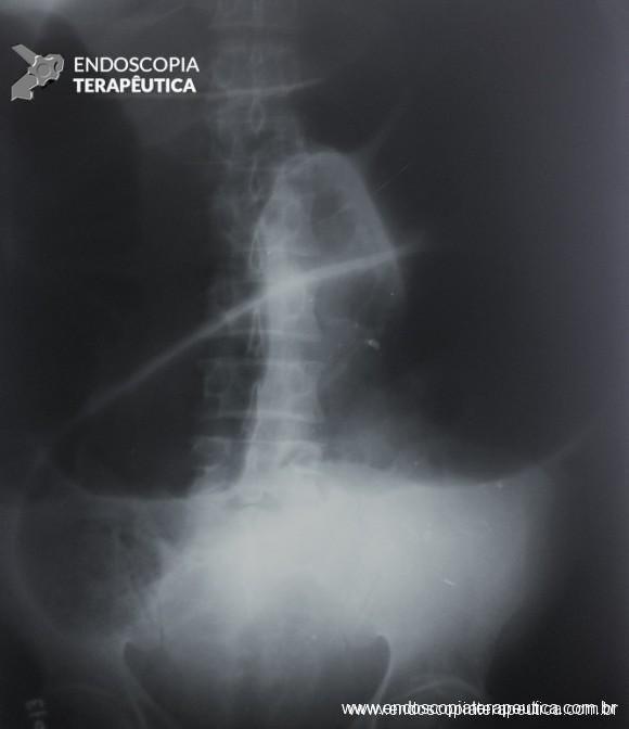
  • In hemodynamically stable patients, an abdominal radiograph aids in the initial evaluation (finding of “coffee bean” and, in patients with incompetent ileocecal valve, small bowel distension). Computed tomography is used to confirm the diagnosis.
Abdominal X-ray showing the “coffee bean” sign, indicative of colon volvulus

Sigmoid Volvulus

  • Hemodynamically stable patients without signs of peritonitis or evidence of perforation should undergo rectosigmoidoscopy to assess the viability of the sigmoid, untwist the torsion, and decompress the colon, effective therapy in 60 to 95% of cases. It is possible to maintain a decompression tube after rectosigmoidoscopy. The recurrence rate is 43 to 75% in cases not subjected to subsequent surgical intervention.
  • Urgent sigmoidectomy is indicated when endoscopic detorsion is unsuccessful and in cases of colon suffering or perforation, as well as in patients with signs of peritonitis or septic shock. After resection of the twisted segment, the decision to perform a primary anastomosis, terminal colostomy, or anastomosis with diversion should be individualized considering the clinical context of the patient at the time of surgery, the conditions of the remaining colon, and comorbidities.
  • Patients who undergo successful endoscopic detorsion are candidates for segmental colectomy during the same hospital admission to prevent recurrent volvulus and its complications. Non-resection operations, including only detorsion, sigmoidopexy, and mesosigmoidoplasty, are inferior to colectomy for the prevention of recurrent volvulus.
  • Endoscopic fixation of the sigmoid may be considered in selected patients in whom surgical intervention is prohibitively risky.

Cecal Volvulus

  • Attempts at endoscopic reduction of cecal volvulus are not recommended.
  • Segmental resection is the treatment of choice for patients with cecal volvulus. Nonviable or ischemic cecum is present in 18% to 44% of patients with cecal volvulus and is associated with a significant mortality rate.
  • In the case of cecal volvulus with viable intestine, the use of non-resection surgical procedures should be limited to patients without clinical conditions for resection.

Acute Colonic Pseudo-Obstruction (Ogilvie’s Syndrome)

  • The initial evaluation should include history and physical examination, laboratory tests, and imaging diagnosis.
    In the absence of fever, leukocytosis, peritonitis, pneumoperitoneum, or cecal diameter > 12 cm, initial therapy consists of correcting hydroelectrolytic disorders, volume replacement, avoiding or minimizing the use of opioids, avoiding anticholinergic medications, and identifying and treating concomitant infections. Deambulation, fasting, positioning maneuvers (knee-chest or prone) to promote intestinal motility, and decompression with nasogastric and rectal tubes are also recommended. Oral osmotic laxatives should be avoided as they can worsen colon dilation. Abdominal radiographs are part of the daily evaluation, accompanied by physical examination.
  • The initial treatment is clinical support and includes the exclusion or correction of conditions that predispose patients to the condition or prolong its course.
  • Pharmacological treatment with neostigmine is indicated when the condition does not resolve with supportive therapy.
  • Endoscopic decompression of the colon should be considered in patients with Ogilvie’s syndrome in whom neostigmine therapy is contraindicated or ineffective.
  • Surgical treatment is recommended in cases complicated by ischemia or perforation of the colon or refractory to pharmacological and endoscopic therapies.

References:

  1. Alavi K, Poylin V, Davids JS, Patel SV, Felder S, Valente MA, Paquette IM, Feingold DL; Prepared on behalf of the Clinical Practice Guidelines Committee of the American Society of Colon and Rectal Surgeons. The American Society of Colon and Rectal Surgeons Clinical Practice Guidelines for the Management of Colonic Volvulus and Acute Colonic Pseudo-Obstruction. Dis Colon Rectum. 2021 Sep 1;64(9):1046-1057. doi: 10.1097/DCR.0000000000002159. PMID: 34016826.
  2. Yeo HL, Lee SW. Colorectal emergencies: review and controversies in the management of large bowel obstruction. J Gastrointest Surg. 2013;17:2007–2012.
  3. Bauman ZM, Evans CH. Volvulus. Surg Clin North Am. 2018;98:973–993.
  4. Quénéhervé L, Dagouat C, Le Rhun M, et al. Outcomes of first-line endoscopic management for patients with sigmoid volvulus. Dig Liver Dis. 2019;51:386–390.

How to cite this article

Camargo MGM. Treatment of Colonic Volvulus and Acute Colonic Pseudo-Obstruction (Ogilvie’s Syndrome). Endoscopy News, 2024, vol1. Available at: https://endoscopy.news/2024/02/05/treatment-of-colonic-volvulus-and-acute-colonic-pseudo-obstruction-ogilvies-syndrome/


ENDOSCOPIC FINDINGS RELATED TO H. PYLORI INFECTION

The Helicobacter pylori

The Helicobacter pylori is a gram-negative bacterium that induces cellular and chemical reactions in the stomach, being considered a human carcinogen. Its diagnosis and treatment play an important role in the prevention of associated diseases, such as gastric cancer, ulcers, MALT lymphoma, and hyperplastic polyps. (To learn more about H. PYLORI, visit this post 1 and this post 2)

There are several diagnostic tests, from non-invasive (serology, breath test, and fecal antigen) to invasive (urease, culture, and histology). Non-invasive methods have high accuracy, but do not assess changes in the gastric mucosa. To learn more about diagnostic tests visit this article.

Due to the focal nature of bacterial colonization, the accuracy of invasive methods depends on the location, number, and size of biopsies. These, when poorly directed, can result in false negatives. Thus, it is important to evaluate the endoscopic predictors of the presence or absence of H. pylori in order to direct biopsies to areas with a higher probability of infection, as well as to avoid them when the positive predictive value is high.

Studies have shown that, although not pathognomonic, some endoscopic findings are associated with the presence of H. pylori. Recently, new chromoendoscopy and magnification technologies have allowed the analysis of the microstructure of the gastric mucosa and, consequently, greater accuracy in determining the infection status (absence, active infection, and post-eradication).

The aim of this article is to assist endoscopists in assessing endoscopic findings related to H. pylori both with white light, as well as chromoendoscopy and magnification.

So let’s go!

2. Findings of the non-infected stomach

2.1 With white light

The normal stomach has a pinkish-red and shiny color, and the mucosal folds are present uniformly. The mucus should be hyaline and often forming a small pool. In the body and fundus, the folds are more concentrated on the greater curvature, in a tent shape and tend to disappear with insufflation. The antrum is flat, with a clear hue.

2.2 With chromoscopy and magnification

To understand the changes visualized in endoscopic magnification, we first need to know the histomorphology of the normal gastric mucosa.

To learn more about the normal histology of the stomach Click here

In summary, the mucosa of the gastric body is composed of cryptic orifice (OC), marginal epithelium of the crypt (MCE), subepithelial capillary network (SECN), collecting vessels, and intervening spaces (between the crypts), as shown in the following scheme:

In the stomach not infected by H. pylori, the subepithelial capillary network is present, regularly throughout the body, called RAC (regular arrangement of collecting venules). The negative predictive value of this finding is greater than 90%, which means that its presence in the distal body’s lesser curvature and incisura is strongly associated with the condition of non-infection by H. pylori.

We can also observe that both the crypt orifice and the marginal epithelium are oval, regular, and symmetrical. The subepithelial capillary network (SECN) is regular and fine, in a honeycomb pattern.

Figure 1: Endoscopic findings of a stomach not infected by H. pylori. (a) The mucosa is smooth, shiny, with uniformly distributed folds. (b) Collecting venules with regular distribution (RAC). (c) RAC in detail with white light and without magnification. (d) Magnification showing the vascular pattern of subepithelial capillaries in “honeycomb”, round crypt orifices (brownish) and marginal epithelium of the crypt in an oval and regular shape. The RAC has a bluish color (cyan).


3. Findings of the infected stomach

3.1- With white light

With white light, the endoscopic findings most associated with H. pylori infection are: diffuse hyperemia, petechial redness (“speckled”) of the fundus and proximal body, thickened and tortuous folds, mucosal edema, fibrinous exudate in the body, and antral nodularity. With the persistence of the infection, there is a decrease in the folds and the submucosal vessels become more visible, findings of atrophic gastritis.

In a prospective multicenter study, the sensitivity and specificity of the endoscopic findings described above were 94.3% and 62.8% (KATO,2013). Diffuse hyperemia was considered the most reliable characteristic by experienced endoscopists.

We know that this infection starts in the antrum and progresses to the body. However, in the antrum, the diagnostic accuracy is lower, as the vessels are located more deeply, hindering their visualization. Therefore, we must first assess the presence or absence of hyperemia in the body. When this assessment is difficult, we should pay attention to petechial redness, edema, folds, and fibrinous exudate.

As previously described, the absence of the regular pattern of collecting venules (RAC negative) may be associated with active infection by H. pylori, but the specificity of this finding is low. In a Brazilian study (Fiuza F, Martins BC, 2021), the absence of RAC was associated with only 50.6% positivity of H. pylori. In other words, the absence of RAC has high accuracy for the presence of the bacterium, but the infection is not always what causes its loss.

It is important to remember that, in H. pylori infection, the RAC initially disappears in the distal body’s lesser curvature and incisura, being these the most specific places to be analyzed. However, in chronic gastritis, when antral atrophy extends precisely through the incisura and lesser curvature of the distal body, there may be disappearance or deformity of the RAC, even in eradicated patients, making its analysis difficult. In these cases, we need to look for the RAC in the distal body mucosa away from atrophy.

Another important fact is that the last region where the RAC is preserved is in the proximal body and gastric fundus, places that are not recommended for evaluating bacterial infection.

Figure 2: Endoscopic findings of a stomach infected by H. pylori. (a) diffuse redness, (b) petechial redness and edema, (c) engorgement of folds, (d) fibrinous exudate in the body, (e) antral nodularity, (f) mucosal edema and absence of RAC, (g) atrophy.

3.2- With magnification

From a histomorphological perspective, with H. pylori infection, the crypts become larger and irregular, surrounded by erythema and grooves. We can no longer see the subepithelial capillary network, as inflammatory cells, edema, degenerated epithelium, and rupture of the microvascular network prevent its adequate visualization. The crypt orifices become asymmetrical and white due to the deposit of inflammatory content inside the glands. As atrophy expands, the marginal epithelium of the crypts becomes enlarged and has an irregular and elongated/curved shape. This is what we call “antralization” of the gastric body.

In a Brazilian study (Fiuza F, Martins BC, 2021), it was found that it is possible to identify these changes in the gastric mucosa (especially the presence or absence of RAC) using near focus technology, since gastroscopes with magnification are not yet widely available.

Figure 3: Endoscopic magnification of a stomach infected by H. pylori: edematous mucosa, with redness and decreased mucosal folding. There was a loss of the normal subepithelial capillary network and collecting venules. The crypts become more elongated and reticular in appearance (“antralization”), with erythema and dilation of subepithelial vessels.

4. Findings of the treated stomach (post-eradication)

It still remains controversial whether H. pylori treatment can reverse atrophic gastritis and intestinal metaplasia. Moreover, it can take up to 10-15 years for the mucosa to recover and return to normal.

After eradication, the non-atrophic areas dissipate the inflammation and the atrophic areas become relatively reddish when compared to the adjacent mucosa. This confers the “map-like” pattern. This pattern may be associated with the development of both primary and metachronous gastric cancer even after effective treatment of H. pylori.

Another characteristic described is the “cracked” pattern, where grooves appear in the antral mucosa, indicating reparative mucosa.

Studies have shown that eradication therapy can alter the characteristics of the repaired stomach, causing difficulty in diagnosing early gastric cancer. That is why the post-eradication status must be distinguished from the negativity of H. pylori.

 Figure 4: Map-like pattern: flat or superficially depressed red lesions of various shapes, sizes, and densities of hyperemia. It is believed that the mechanism of the map-like appearance is the strengthening of the contrast between the non-atrophic mucosa and the atrophic mucosa after the disappearance of diffuse redness. This finding is not always present, however, when observed, it is indicative of gastric mucosa post eradication.
Figure 5: Atrophy pattern with “cracks” (Cracked pattern). This finding

References

  1. Anagnostopoulos GK, Yao K, Kaye P, Fogden E, Fortun P, Shonde A, Foley S, Sunil S, Atherton JJ, Hawkey C, Ragunath K. High-resolution magnification endoscopy can reliably identify normal gastric mucosa, Helicobacter pylori-associated gastritis, and gastric atrophy. Endoscopy. 2007 Mar;39(3):202-7.
  2. Yao K. The endoscopic diagnosis of early gastric cancer. Ann Gastroenterol. 2013;26(1):11-22. 
  3. Yagi K, Nakamura A, Sekine A. Comparison between magnifying endoscopy and histological, culture and urease test findings from the gastric mucosa of the corpus. Endoscopy. 2002 May;34(5):376-81. 
  4. Yuan C, Lin XM, Ou Y, Cai L, Cheng Q, Zhou P, Liao J. Association between regular arrangement of collecting venules and Helicobacter pylori status in routine endoscopy. BMC Gastroenterol. 2021 Oct 20;21(1):389. 
  5. Glover B, Teare J, Patel N. A systematic review of the role of non-magnified endoscopy for the assessment of H. pylori infection. Endosc Int Open. 2020 Feb;8(2):E105-E114.
  6. Qi Q, Guo C, Ji R, Li Z, Zuo X, Li Y. Diagnostic Performance of Magnifying Endoscopy for Helicobacter pylori Infection: A Meta-Analysis. PLoS One. 2016 Dec 19;11(12):e0168201.
  7. Weng CY, Xu JL, Sun SP, Wang KJ, Lv B. Helicobacter pylori eradication: Exploring its impacts on the gastric mucosa. World J Gastroenterol. 2021 Aug 21;27(31):5152-5170.  
  8. Nishikawa Y, Ikeda Y, Murakami H, et al. Classification of atrophic mucosal patterns on Blue LASER Imaging for endoscopic diagnosis of Helicobacter pylori-related gastritis: A retrospective, observational study. PLoS One. 2018;13(3):e0193197.
  9. Toyoshima O, Nishizawa T, Koike K. Endoscopic Kyoto classification of Helicobacter pylori infection and gastric cancer risk diagnosis. World J Gastroenterol. 2020 Feb 7;26(5):466-477.
  10. Ono S, Dohi O, Yagi N, Sanomura Y, Tanaka S, Naito Y, Sakamoto N, Kato M. Accuracies of Endoscopic Diagnosis of Helicobacter pylori-Gastritis: Multicenter Prospective Study Using White Light Imaging and Linked Color Imaging. Digestion. 2020;101(5):624-630.
  11. Kato T, Yagi N, Kamada T, Shimbo T, Watanabe H, Ida K; Study Group for Establishing Endoscopic Diagnosis of Chronic Gastritis. Diagnosis of Helicobacter pylori infection in gastric mucosa by endoscopic features: a multicenter prospective study. Dig Endosc. 2013 Sep;25(5):508-18.
  12. Fiuza F, Maluf-Filho F, Ide E, Furuya CK Jr, Fylyk SN, Ruas JN, Stabach L, Araujo GA, Matuguma SE, Uemura RS, Sakai CM, Yamazaki K, Ueda SS, Sakai P, Martins BC. Association between mucosal surface pattern under near focus technology and Helicobacter pylori infection. World J Gastrointest Endosc. 2021 Oct 16;13(10):518-528.

How to cite this article

Nobre R, Baba E. ENDOSCOPIC FINDINGS RELATED TO H. PYLORI INFECTION. Endoscopy News; 2024, vol1. Available on: https://endoscopy.news/2024/01/27/endoscopic-findings-related-to-h-pylori-infection


Hereditary Gastric Cancer

Authors: Marcus Fernando Kodama Pertille Ramos and Ítalo Beltrão Pereira Simões

The majority of gastric cancer (GC) cases are sporadic, but about 10% present familial aggregation and 1 to 3% have a hereditary cause. Knowledge of hereditary syndromes as a causal factor for colorectal cancer (CRC) is well estabilished, but for GC this is less publicized, which can impair early diagnosis and proper follow-up.

Hereditary GC can occur with or without the presence of polyposis, as in the case of Hereditary Diffuse Gastric Cancer, Li-Fraumeni, BRCA1, BRCA2, and Lynch syndromes. In this article, we will briefly describe the main hereditary GC syndromes associated with polyposis, leaving those not associated for a future article.

1. HEREDITARY GC ASSOCIATED WITH POLYPOSIS

1.1 FAMILIAL ADENOMATOUS POLYPOSIS (FAP)

FAP results from a mutation in the APC tumor suppressor gene causing a very high risk of CRC.

About 51 to 88% of patients have gastric polyps, mainly fundic gland polyps (FGP). The incidence is high even in attenuated FAP. They tend to be numerous, and the term gastric polyposis can be used only when more than 20 are present.

Low-grade dysplasia may be present in up to 44% of fundic gland polyps. Adenomatous polyps are detected in about 20% of patients with FAP.

Upper endoscopic screening is recommended at the time of colonic polyposis manifestation or from the age of 25. The interval of performance will depend on the findings and also according to the need for follow-up of papillary adenoma, when present, according to the Spigelman score.

Gastric polyposis in a patient with FAP. Images show numerous polyps in the cardia, fundus, body, and antrum.

1.2 PEUTZ-JEGHERS SYNDROME (PJS)

PJS is an autosomal dominant disorder, characterized by the development of gastrointestinal hamartomatous polyposis mainly in the jejunum associated with the presence of melanocytic macules.

The clinical diagnosis is based on the confirmation of the presence of hamartomatous polyps associated with a positive family history and hyperpigmentation of mucous membranes, fingers, and external genitalia.

Gastric polyps are detected in 25% of cases, compared with 70-90% found in the small intestine and 50% in the colon. The morphological appearance of the gastric polyp in PJS resembles a villous pattern of hyperplastic epithelial proliferation, making it difficult to distinguish from juvenile and hyperplastic polyps.

Dysplasia is rarely detected in the polyps, but individuals with PJS have a 29% risk of developing GC, mainly of the intestinal type.

Screening should be started early in childhood with initial endoscopy, with frequency depending on the findings. From the age of 50, the risk of GC increases and the frequency should be more frequent between 1 to 2 years.

Image sequence showing a polyp in the gastric body region in a patient with Peutz-Jeghers Syndrome, lower images with image magnification.

1.3 JUVENILE POLYPOSIS SYNDROME

An autosomal dominant syndrome that leads to the development of polyps throughout the gastrointestinal tract mainly in the colon and rectum.

Criteria for clinical suspicion of the syndrome include more than 5 colorectal juvenile polyps, juvenile polyps throughout the gastrointestinal tract, or more than 1 juvenile polyp with a positive family history. The definitive diagnosis is made from one of the clinical suspicion criteria in the presence of the BMPR1A and SMAD4 genes in the genetic test.

Juvenile polyps are hamartomatous polyps that develop from normal tissue of the gastrointestinal tract. The usual endoscopic appearance is of a pedunculated, multilobed, soft polyp ranging from small polyps to giant polyps. In up to 75% of cases, other types of polyps are present together. Severe gastric polyposis can occur causing anemia, hematemesis, protein-losing enteropathy, and obstructive symptoms. Progression to GC occurs in up to 21% of cases with an average age of 58 years.

Endoscopic screening is recommended from adolescence with annual endoscopies.

Learn more about juvenile polyposis by clicking this post

Upper images show a hamartomatous juvenile polyp in the cardia region in a patient with juvenile polyposis. Lower images show the different aspects and sizes of the polyps found in the syndrome.

1.4 MUTYH-ASSOCIATED POLYPOSIS (MAP)

MAP is a rare syndrome, autosomal recessive, associated with mutation in the MUTYH gene that participates in DNA repair processes. Patients with MAP are predisposed to CRC, breast, and ovarian cancer.

Gastric polyps are detected in about 10 to 33% of cases and the majority are adenomas and FGP.

The risk of GC is low (2%) but occurs in younger patients (median age of 38 years). On the other hand, the risk of duodenal cancer is high and can occur in 17% of cases.

1.5 GASTRIC ADENOCARCINOMA WITH PROXIMAL POLYPOSIS (GAPPS)

This syndrome is characterized by the development of proximal gastric polyposis including the fundus and body forming a carpet of small polyps usually smaller than 1 cm. The histological type of the polyps is varied and can be FGP, hyperplastic, adenomas, and mixed.

The criteria for clinical diagnosis include detection of more than 100 polyps or more than 30 polyps with a positive family history in a first-degree relative, polyps restricted to the body and fundus without the presence of colorectal polyps, morphology of FGP with areas of dysplasia or carcinoma, exclusion of other syndromes, and use of proton pump inhibitors.

Case series have reported an incidence of 12.7% of GC, all of the intestinal type.

Endoscopic follow-up should be performed, but in cases with multiple polyps, the evaluation of polyps with signs of degeneration may be impaired, indicating total gastrectomy.

Images of GAPPS showing involvement of the body and fundus with progressive decrease in the number of polyps in the distal stomach.
Table summarizing the main characteristics of genetic syndromes related to hereditary gastric cancer, dividing them into those associated and not associated with polyposis of the gastrointestinal tract.

References

  1. Clauditz TS, Moore M, Setia N, et al. Syndromic gastric polyposis and hereditary gastric cancers. Diagnostic Histopathologic 2019; 26(1):39-46.
  2. Mahon SM. Hereditary Polyposis Syndromes. Gentics and Genomics 2018; 22(2): 151-6
  3. Cardoso DM. Síndromes de polipose colorretal. Endoscopia Terapêutica; 2020. Disponível em: http://endoscopiaterapeutica.com.br/assuntosgerais/sindromes-de-polipose-colorretal/

How to cite this article

Kodama, MFKP and Simoes IBP. Hereditary gastric cancer. Gastropedia 2022. Available at: https://endoscopiaterapeutica.com.br/assuntosgerais/cancer-gastrico-hereditario


Well-performed endoscopic piecemeal mucosal resection (EPMR) is better than a failed attempt of en-bloc mucosectomy (EMR) – Tips for performing without fear!

A real-life case we encounter during colonoscopy schedules:

A 60-year-old man, previously healthy, with no comorbidities or relevant family history for gastrointestinal tract neoplasms, underwent his first colonoscopy for colorectal cancer screening/prevention and obtained the following finding:

– Laterally spreading tumor of the granular type, homogeneous (LST-G-H), with regular surface and vasculature on virtual chromoendoscopy with NBI, measuring about 4cm, located in the cecum (Paris 0-IIa / JNET 2A).

Laterally spreading tumor, granular type, homogeneous (LST-G-H) of the cecum measuring 4cm
Regular surface with dendritic pattern
Regular and uniform distribution of vessels with a well-ordered reticular pattern

Faced with a lesion of this size, it is natural that doubts arise about the ideal treatment to be offered to the patient, especially in those who do not master advanced endoscopic resection techniques, particularly Endoscopic Submucosal Dissection (ESD).

Classification of LSTs

  • The term LST was originally proposed by Kudo et al and describes a pattern of growth of colorectal tumors with a diameter >10 mm, which tend to spread laterally with a smaller vertical axis.
  • LSTs can be classified into two types: granular type LST (LST-G), with granules and nodules on the tumor surface, and non-granular type LST (LST-NG), with a flat and smooth surface.
    • The former can be subclassified: homogeneous granular type LST (LST-G-H), with granules or nodules uniformly distributed on the tumor surface, and mixed nodular type LST (LST-G-M), with nodules of different sizes on the surface;
    • the latter is also subclassified: pseudo-depressed non-granular type LST (LST-NG-PD), when there is a depression in the center of the lesion, and flat-elevated non-granular type LST (LST-NG-F), without depression, that is, completely flat.

learn more about macroscopic classification in this post: click here

Prevalence and location of LSTs

  • The estimated frequency of LSTs among all epithelial colorectal tumors, excluding advanced carcinomas, varies in the literature, averaging 4.5%, however, more recent studies with larger case series point to a prevalence that can reach up to 12.3%.
  • LST-Gs are the most prevalent, accounting for about 65-70% of all LSTs. While LST-G-Hs tend to be located more in the proximal colon, LST-G-Ms affect the rectum more often and usually reach larger diameters. LST-NGs are more commonly found in the transverse colon.

LST x risk of malignancy

  • Each subtype has different and unique characteristics, which requires a decision on the treatment policy to be individualized.
  • More than half of LST-G-H lesions are adenomas, with the presence of carcinoma being rare, even when they reach a large diameter, however, even when present, it tends to be restricted to the mucosa (Tis – intramucosal).
  • As for LST-NGs, although both LST-NG-F and LST-NG-PD are considered as subtypes, the latter has a higher rate of submucosal invasion even at small sizes, whose nature tends to be multifocal, and it is the subtype with the highest risk among all LSTs. LST-NG-Fs have a low risk of submucosal invasion, and in the few cases present, it tends to be focal.

LST x choice of endoscopic treatment

  • ESD is widely used in the upper gastrointestinal tract, however, due to anatomical and histological differences between the colon and the stomach, it is not established as the standard therapeutic technique for colorectal tumors. Furthermore, it is essential to consider, even despite advances in recent years, that the ESD technique is still quite scarce in most medical centers, so the choice of endoscopic treatment technique cannot cause more difficulties than the problem itself.
  • Almost all LST-G-Hs do not invade the submucosa. Despite being higher compared to ESD, when well executed, the recurrence rate after EPMR is low, without clinically relevant concerns, since when it occurs it is usually unifocal, small and easily treated in a single session. Therefore, in this morphological type, EMR and EPMR can be adopted as the first option, especially for their higher safety profile.
  • Because LST-NG-PDs have the highest rate of submucosal invasion, even at small sizes, with a more multifocal invasive nature and a tendency for greater depth, regardless of whether it is observed only in one location, en bloc resection, mainly by ESD, should always be considered the first option to allow a more reliable pathological evaluation.
  • As for LST-G-Ms, most submucosal invasions occur below the largest nodule, however, in up to 17.1% of them, there are also foci of invasion outside the dominant nodule (multifocal invasion). As this morphological type reaches the largest diameters, en bloc resection by EMR is often considered difficult. For these reasons, when opting to perform EPMR, it is necessary to ensure the resection of the dominant nodule in a single piece or proceed with ESD, in order to obtain accurate pathological diagnoses. It is worth remembering that large LST-G-Ms are more frequently located in the rectum, where both the safety profile and the outcomes of a non-curative endoscopic treatment due to the impossibility of histological evaluation (definitive colostomy) favor the performance of ESD over EPMR.
  • LST-NG-Fs have a much lower risk of submucosal invasion compared to LST-NG-PDs, in some series even comparable to the risk of LST-G-Hs, therefore, several of those lesions can be cured by endoscopic treatment with EMR or EPMR. However, as the size of this morphological type is associated with a greater possibility of submucosal invasion, especially when larger than 30 mm in diameter, en bloc resection by ESD can also be adopted if they are difficult to remove en bloc by EMR.

Golden tips for performing an EPMR

1. Spend enough time evaluating the lesion to be treated.

Make sure to inspect the lesion to be resected. Make a point of spending enough time evaluating the morphology of the lesion according to the Paris classification, as well as the vascular and glandular patterns. It’s not a waste of time, in fact, you’ll save time by deciding the best way to approach the lesion! Pay attention to the margins, as they may extend beyond the fold. Inspect the lesion with high-definition white light and conventional or virtual chromoendoscopy. A thorough evaluation can identify lesions with possible submucosal invasion and consequently those patients who will benefit from en bloc resection.

2. Do not underestimate the relevance of the lesion’s position.

Have a good position with the device straightened and relaxed. Position the lesion between 5 and 6 o’clock in the endoscopic field. The device and accessory should respond “one to one” to the movements of the hands, fingers, and also the wheels. Working in the best position is extremely effective in minimizing risks and maximizing the outcome of the resection. If a variable stiffness endoscope is being used, take advantage of the potential for retroflexion of the tip. Position the patient so that any fluid or resected pieces accumulate away from the lesion, so that the work field is kept clean and the ideal vision is preserved in case of complication.

3. Choose the snare wisely.

Depending on the morphology or size of the polyp, selecting the most suitable snare can make a difference in the success of the procedure and, therefore, in the outcomes. Small (10–20 mm) or large (25–33 mm) rigid snares with braided wire should be preferred for EPMR and en bloc EMR, respectively. On the other hand, monofilament snares may be the best option for capturing lesions that have difficulty lifting, such as recurrence after EMR or situations where there has been a previous attempt at resection. Use the device as an extension of your hand, placing it parallel to the wall. Adapt the cut to the plane of the lesion, fragment by fragment. The more angle you create between the snare and the wall, the greater the likelihood of involving the muscularis propria. Close the snare well to keep the lesion in place before resecting it. Be aware of the possibility of submucosal fibrosis resulting from previous biopsy collections, previous resection attempts, and LST-NG, as in these situations snare apprehension can be difficult, eventually requiring alternative techniques for lesion removal.

4. Don’t be greedy!

Once the EPMR technique is chosen, keep in mind that the goal should be the complete removal of the lesion with the highest possible safety. For this, the correct strategy is fundamental: do not make the submucosal bubble all at once, instead, make successive injections followed by cutting, preferably in the proximal-distal direction; whenever available, prefer viscous solutions, which ensure greater patency of the bubble; use a smaller snare (10-15 mm) to grasp the formulated bubble, in addition to facilitating, it reduces the risk of perforation when trying to inadvertently grasp the entire lesion.

5. Do not panic with bleeding.

When intraprocedure bleeding (IPB) occurs, do not panic – it’s just bleeding. Although it is true that only practical experience can make you confident in the face of IPB, be prepared to approach it systematically, as you would with any other endoscopic procedure. Before starting the procedure, you must ensure that your endoscopy set is fully equipped and capable of dealing with all types of IPB. Make conscious use of everything you can, without panicking. Use the irrigation pump to remove blood from the target tissue and clean the point where you need to intervene. If you judge that the vessel is small (up to about 2 mm), you can coagulate it immediately with the tip of the snare in “soft coagulation” mode. On the other hand, if the vessel is larger than 2 mm, the use of a coagulation forceps is a more effective strategy. While waiting for the accessory, if you are using a cap attached to the tip of the device, use it as a “finger” and press on the vessel. When you are ready with your device, use the water pump again to clean the area, open the forceps and grasp the vessel, pulling it towards you (and away from the wall) before coagulation. The use of argon plasma coagulation (APC) during EMR should be minimized, as well as hemostatic clips, which should be used when you have tried everything and the bleeding continues.

Given the above, for the case exemplified above, it was decided to proceed with resection using the EPMR technique, both for the morphological type (LST-G-H) and characteristics of the lesion surface (JNET 2A), as well as for the location (cecum), which presents a higher risk of complications.

References

  1. Papparella L et al. Efficacy and safety of endoscopic resection techniques of large colorectal lesions: experience of a referral center in Italy. Eur J Gastroenterol Hepatol 2022; 34: 375–381.
  2. Ishigaki T et al. Treatment policy for colonic laterally spreading tumors based on each clinicopathologic feature of 4 subtypes: actual status of pseudo-depressed type. Gastrointest Endosc 2020; 92: 1083-94.
  3. Auriemma F and Repici A. Mistakes in endoscopic resection and how to avoid them. UEG Education 201


Serrated Polyposis Syndrome (SPS)

The sessile serrated lesion (SSL) is the new term for lesions previously called sessile serrated adenoma (SSA) and SSL with dysplasia is the term used for lesions previously called SSA with dysplasia. A detailed review of serrated lesions can be found in this other article.

Based on this, Serrated Polyposis Syndrome (SPS) is a rare condition characterized by multiple colorectal serrated lesions and increased risk of colorectal cancer.

  • The prevalence varies from 0.03% to 0.5% in patients undergoing colonoscopy.
  • The diagnosis usually occurs between 50-55 years, although it ranges from 20-70 years.
  • Similar prevalence in men and women.

Click here to learn about other colorectal polyposis syndromes.

Updated Criteria for Serrated Polyposis:

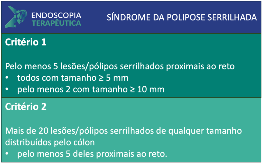
Compared to the previous edition, the WHO in 2019 updated the diagnostic criteria, keeping only 2 of the 3 clinical criteria for the definition of SPS:

  1. Criterion: At least 5 serrated lesions/polyps proximal to the rectum, all measuring ? 5 mm, with at least 2 measuring ? 10 mm.
  2. Criterion: More than 20 serrated lesions/polyps of any size distributed throughout the colon, with at least 5 of them proximal to the rectum.
  • Patients who meet at least one criterion are diagnosed with serrated polyposis.
  • All subtypes of serrated polyps (sessile serrated lesion, hyperplastic polyp, traditional serrated adenoma, unclassified serrated adenoma) are included in the count.
  • The count is cumulative over several colonoscopies.
  • Studies report a cancer risk between 15–30% in patients with SPS, varying according to age, polyp phenotype, and high-risk histological features.
  • Rigorous endoscopic surveillance (annual colonoscopy) is recommended.
  • More recent studies have shown that most patients have endoscopic control with a reduction in the number and size of polyps after 2-3 annual colonoscopies, suggesting spacing the surveillance interval to 2 years after this initial stage [1].

Summary of Changes:

Diagnostic criteria for Serrated Polyposis Syndrome according to WHO 2019

Reference

  1. MacPhail M.E., Thygesen S.B., Patel N., Broadley H.M., Rex D.K. Endoscopic control of polyp burden and expansion of surveillance intervals in serrated polyposis syndrome. Gastrointest. Endosc. 2019;90:96–100. doi: 10.1016/j.gie.2018.11.016. 

How to cite this article

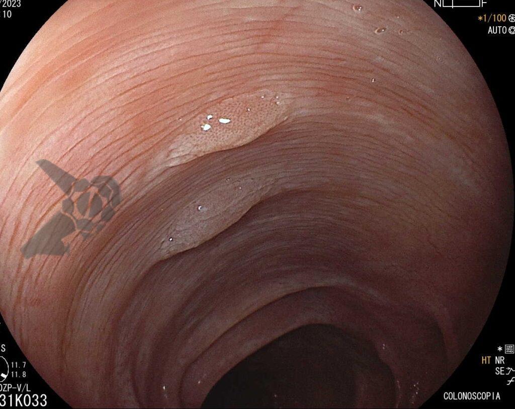
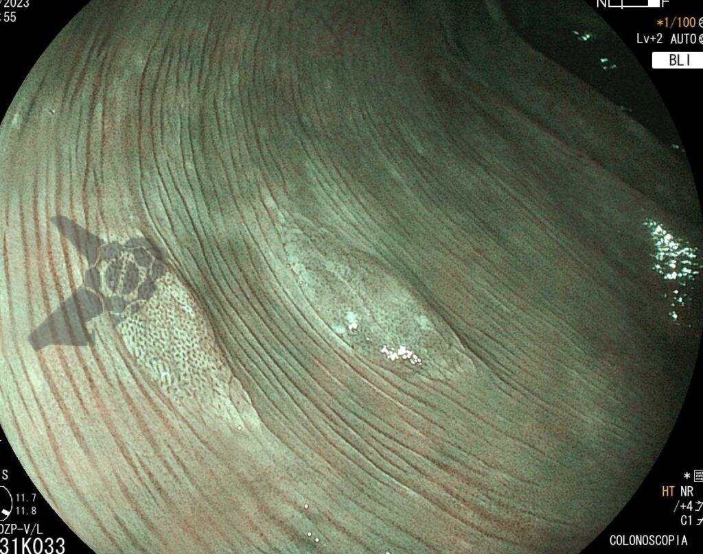
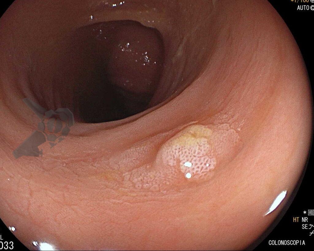
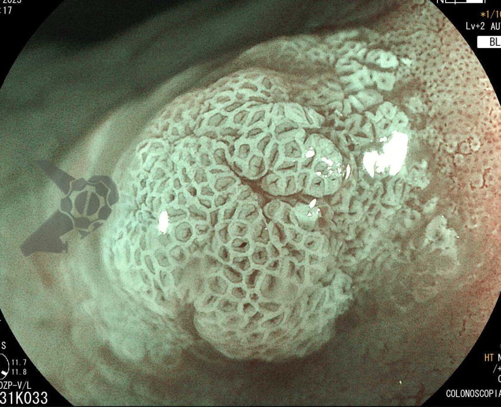
Martins BC and Tanigawa R. Serrated Polyposis Syndrome (SPS). Endoscopia Terapeutica, 2024 vol. 1. Available at: https://endoscopiaterapeutica.com.br/assuntosgerais/sindrome-de-polipose-serrilhada-sps/